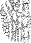
Dicotyledon Leaf
A: "Camera-lucida drawing of a bleached leaf of a Dicotyledon, showing the course of the vascular bundles,…
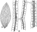
Monocotyledon Leaf
B: "Camera-lucida drawing of a bleached leaf of... a Monocotyledon, showing the anastomosis of the parallel…
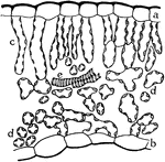
Leaf Water Flow
"Semi-diagrammatic cross section of a leaf showing by arrows how the water passes from the tracheal…
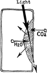
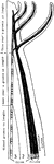
Leaf Water Flow
"Diagram to show the path of the water as it rises to, and escapes from, the leaves." -Stevens, 1916
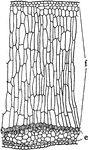
Yellow Poplar Stem Tissue
The cross section of the stem of yellow poplar, which carries large amounts of water.
Water Cress Stem Tissue
The cross section of the stem of water cress, which carries very little water.
P. Galapageium Stem Tissue
The cross section of the stem of Pisdium Galapageium, which carries very little water.
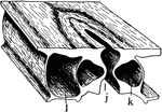
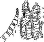
Stoma
"A typical stoma in cross section and surface view combined. k, guard cell; j, the gap or stoma between…
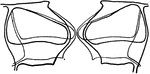
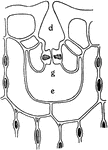
Stoma Guard Cells
"A, diagram showing relative position of the guard cells in cross section in the open and closed positions;…
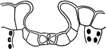
Stomata Formation
"B and C, early stages in the formation of stomata; at s, mother cells of guard cells are shown. D,…
Guard Cell Experiment
"Diagram of apparatus showing how the guard cells draw apart; j, j, position of the rubber tubing when…
Depressed Stoma
"A, depressed stoma of the under side of a leaf of Amherstia nobilis." -Stevens, 1916
Depressed Stoma
"B, depressed stoma of Hakea suaveolens. g stands beneath the guard cells; d, outer, and e inner, cavities."…
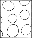
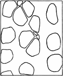
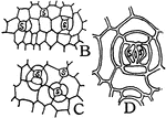
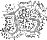
Intercellular Spaces of a Plant
"Diagram suggestive of the distribution of intercellular spaces throughout a plant. The heavy horizontal…
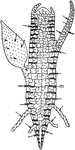
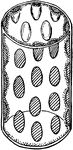
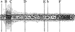
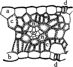
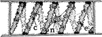
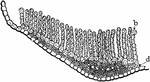
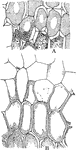
Juncus Stem
"A, cross section of stem of Juncus...the black lines traversing the section are really chains of cells…
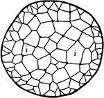
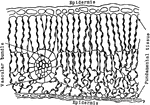
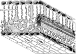
Leaf Epidermis
"Cross section of a portion of the blade of a leaf, showing upper epidermis at a, lower epidermis at…
Palisade Cell
"Diagrammatic representation of a single palisade cell, with chloroplasts lining the walls." -Stevens,…
Euphorbia Palisade Cells
"Starch grains from the palisade cells of a Euphorbia leaf." -Stevens, 1916

Palisade Cell Intake
"Diagram to show the intake of carbon dioxide by the palisade cells from the intercellular spaces, the…
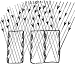
Plant Light Intake
"Diagram showing how the position of the chlorplasts against the vertical walls of the palisade cells…
Palisade Cell
"Diagram to show the activities going on in a palisade cell. The arrows from the chloroplasts into the…
Photosynthesis
"Diagram to show the effect of different portions of the spectrum on photosynthesis. a to F, different…
Intercellular Spaces of Leaf
"Showing intercellular spaces: f, between the palisade cells; e, in a leaf; g, border parenchyma; h,…
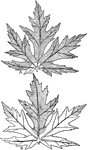
Leaf Architecture
"Diagram to show the architecture of a typical leaf in the region of one of the lateral veins. The shaded…
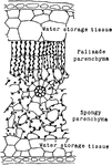
Rubber Leaf Cells
"Cross section through a portion of rubber leaf, showing the large percentage of water-storage tissues…
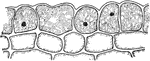
Indian Corn Leaf Cells
"Cross section of a portion of a leaf of Indian corn. a, upper and b, lower epidermis; c, c, palisade…
Codonanthe Leaf Tissues
"Cross section of a portion of leaf of Codonanthe, showing the water-storage tissue at f, and the chlorophyll-bearing…
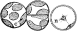
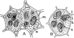
Spirogyra Chloroplast
"B, cell of Spirogyra, with spiral chloroplast at c, nucleus at n, and pyrenoid at e." -Stevens, 1916
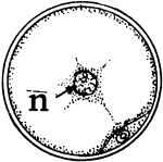
Spirogyra Chloroplast
"C, cross section of Spirogyra cell, with nucleus at n, and section of chloroplast and pyrenoid below."…
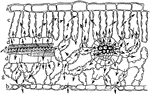
Marchantia Thallus
"Cross section through the thallus of Marchantia. j, stoma leading into a relatively large air-chamber…
Sphagnum Leaf
"Portion of leaf of Sphagnum, in cross section on the left, and surface view on the right. h, hole through…
P. Commune Leaf
"Cross section through a portion of leaf of Polytrichum commune. b, chains of chlorophyll-bearing cells;…
Moss Leaf Chloroplasts
"Cross section, A, and surface view, B, of a leaf of common moss, showing chloroplasts, c." -Stevens,…
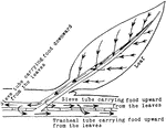
Stored Food
"Diagram to show path of stored food upward through the tracheal tubes, and through the phloem portion…
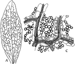
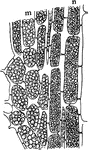
Yellow Poplar Wood
"Showing pitted connections between medullary rays and xylem parenchyma, and between contiguous xylem…
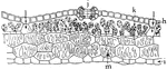
Oak Wood
"Outline of tangential section of wood of oak, to show frequency of medullary rays. The section is 1…
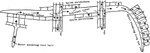
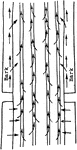
Plant Vein
"Diagram indicating the succession of the conducting tissues of a vein from the base toward the apex.…
Cut Leaf Veins
"Showing the effect of cutting across the veins on the removal of food from the leaf. A, all of the…
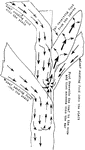
Leaf Food Circulation
"Diagram illustrating the descent of food from the leaf into the stem, and its circulation upward and…
Indian Corn Food Circulation
"Diagram showing how, in Indian corn, the food from the upper and lower leaves finds its way into the…
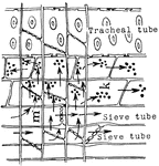
Sieve Tube Food Transportation
"Diagram showing the transport of food through the sieve tubes, medullary rays and tracheal tubes, and…
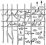
Food Tissues
"Diagram to show the relation of the food-conducting tissues of the leaf to those of the stem; and in…
Grapevine Wood
"Tangential section through the wood of grapevine. m, cells of medullary ray, and n, of xylem parenchyma,…
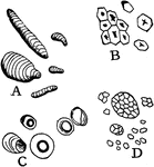
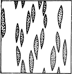
Starch
"Starch from different sources. A, curcuma starch; B, corn starch; C, tapioca starch; D, rice starch,…
Sphaero-Crystals
"Sphaero-crystals of unilin from tuber of Dahlia variabilis. A, precipitated from an aqueous solution;…
I. Balsamina Storage Tissues
"Storage tissues of the cotyledon of Impatiens Balsamina. A, from the resting seed, and B, from a germinating…
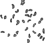
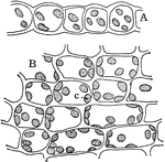
Aleurone Grains
"To show aleurone grains. A, cells from cotyledon of seed of garden bean; n, aleurone grains; m, starch;…
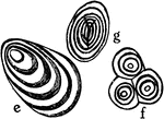
Starch Grain Striations
"Showing concentric and eccentric striations of starch grains. e, potato starch eccentrically striated;…
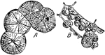
L. Tulipifera Wood
"Tangential section of wood of Liriodendron tulipifera, showing frequency of contact of medullary rays…
Plant Food Storage
"Diagram to show food from the leaves descending through the sieve tubes and being stored in the medullary…
Plant Food Digestion
"The digestion of the stored food and its ascent through the tracheal tubes when growth is resumed in…
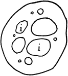
D. Rotundifolia Digestive Gland
"Longitudinal section through a digestive gland of Drosera rotundifolia." -Stevens, 1916
M. Forskalli Leaf
"Cross section of leaf of Mesembryanthemum Forskalii showing a large part of the leaf devoted to the…